For centuries, physicians viewed the heart as a remarkable yet simple machine. It functions as a muscular pump that moves blood through the body. Yet a growing body of research suggests this view is incomplete. According to a new review in the Journal of Taibah University Medical Sciences, the heart may process information. It may also produce hormones linked to bonding and calm. It sends signals that shape how we feel, perceive, and decide.
The review, authored by Dr. Ahmed S. BaHammam of King Saud University, examines whether contemporary cardiac science has moved back toward an older understanding of the heart. The Qur’an and Prophetic tradition, he notes, describe the heart as a center of reasoning, faith, emotion, and moral responsibility. Arab-Islamic scholars, including al-Balkhi, al-Razi, Ibn Sina, and al-Ghazali, linked cardiac physiology to emotion, cognition, and spiritual life. It predated Cartesian physiology by centuries.
Crucially, the review does not claim that science proves scripture. Instead, it shows that independent scientific disciplines have arrived at a portrait of the heart. These fields work without reference to religious texts and align with descriptions in Islamic tradition. In other words, the convergence is suggestive, not conclusive.
A Nervous System Inside the Heart
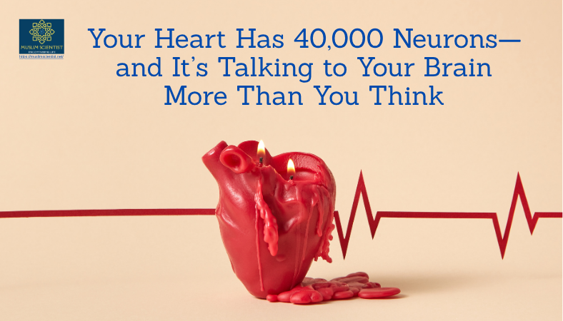
Perhaps the most striking finding comes from neurocardiology. The human heart contains an intrinsic cardiac nervous system (ICNS) of roughly 14,000 to 94,000 neurons. These neurons group into gangliated plexuses on the heart’s surface. They include sensory neurons, processing interneurons, and motor neurons. These elements support local information processing.
This network coordinates beat-to-beat cardiac function through rapid reflexes, with little reliance on the central nervous system. Transplanted hearts, entirely denervated, still exhibit autonomous neural regulation through their intrinsic ganglia. As the review puts it, the ICNS is capable of reflex control of regional cardiac function. This occurs in the absence of all higher elements.
Recent molecular research has shown that these cardiac neurons are not a uniform population. Researchers used single-cell RNA sequencing in mice to find two ICNS neuron subtypes. One subtype supports baseline heart function, while the other may guard against stress-induced sudden death. The investigators selectively ablated the stress-protective subtype. 63% of the animals died from sudden cardiac arrest within minutes of stress exposure. Activating these neurons prevented such deaths. If confirmed in humans, these findings could have significant implications for sudden death prevention.
However, the heart’s nervous system is not as versatile as the brain’s. Brain neurons employ synaptic plasticity extensively for learning and memory, whereas ICNS neurons prioritise stability and direct cardiac control. Nevertheless, the parallel is closer than anyone anticipated a generation ago.
The Heart Speaks to the Brain
The heart communicates with the brain at least as much as the brain communicates with the heart. These findings apply to specific contexts that have been experimentally examined. Approximately 80% of vagal cardiac fibers carry signals from the heart to the brain. Additionally, these signals contribute to emotional and perceptual processing, alongside descending central regulation.
A computational model of brain-heart interplay was applied to EEG and ECG signals from 62 healthy subjects during stimulation. Cardiac sympathovagal activity was found to play a leading and causal role in initiating the emotional response. Ascending vagal modulations preceded neural dynamics and correlated with the reported level of emotional arousal. The heart’s physiological response occurred first and then shaped the brain’s processing of the emotional experience.
This is not the classical model where the brain decides an emotion and then instructs the heart to race. Instead, the heart’s own rhythmic activity initiates and shapes the emotional cascade, which then unfolds through ongoing heart-brain coupling rather than through the heart in isolation.
The clinical consequences of this bidirectional axis can be dramatic. Takotsubo cardiomyopathy, sometimes called “broken heart syndrome,” illustrates how emotional processing in the brain can physically damage the heart. An acute emotional shock, such as grief, terror, or neurological catastrophe, triggers a catecholamine surge. It stuns the left ventricular apex, producing a ballooning pattern on echocardiography. Takotsubo syndrome has been described explicitly as “a disorder of the heart-brain axis.”
Hormones of Bonding and Tranquility
The heart also functions as an endocrine organ. In the late 1990s, Gutkowska et al. demonstrated the presence of functional oxytocin receptors in cardiac tissue. Jankowski et al. found that the rat heart synthesizes oxytocin. Oxytocin mRNA was detected in all chambers, with the highest peptide concentrations in the right atrium. The cardiac oxytocin/oxytocin receptor system was subsequently characterized in human tissue.
Oxytocin is widely recognized as the hormone of social bonding, surging during childbirth, breastfeeding, and close human contact. Cardiac oxytocin slows the heart rate, lowers blood pressure, and promotes parasympathetic tone, while also exerting cardioprotective effects such as reducing infarct size after ischemia and activating anti-inflammatory pathways.
When oxytocin activates its receptors in the atria, a primary downstream effect is the release of atrial natriuretic peptide (ANP). ANP receptors are found in brain regions governing emotional states, including the amygdala and locus coeruleus, the brain’s central norepinephrine hub. Intravenous ANP administration blocked experimentally induced panic attacks in patients with both panic disorder and healthy controls. ANP inhibited the hypothalamic-pituitary-adrenal (HPA) stress axis, reducing cortisol and adrenocorticotropic hormone, while simultaneously dampening sympathetic activation.
Thus, a feedback loop emerges: social bonding raises oxytocin, which triggers cardiac ANP release that acts on the brain to produce anxiolysis and suppress the stress response. Within this loop, the heart functions as an active endocrine node, releasing peptides that modulate central autonomic and stress-regulatory circuits, rather than serving as a passive downstream target.
The Heart Shapes What We Perceive
Neuroscience research now suggests that the heart shapes what the brain perceives, feels, and knows through a process called “cardioception.” Every heartbeat sends a signal to the brain. When the ventricles contract during systole, baroreceptors in the aortic arch and carotid sinus fire, sending ascending signals through the vagus nerve to the brainstem, and from there to the thalamus, insula, and cortex.
The cortical imprint of this process is a measurable electrical response time-locked to each heartbeat, known as the heartbeat-evoked potential (HEP). Source localization and intracranial recordings have traced the HEP to the insula, anterior cingulate cortex, and prefrontal regions. Direct intracranial EEG in patients confirmed that heartbeat-evoked responses arose primarily from the insular and opercular cortices, where the HEP amplitude was modulated during experimental manipulations of bodily self-consciousness.
Does this cardiac monitoring affect what we consciously experience? The evidence is increasingly affirmative. Fearful faces presented during cardiac systole (when baroreceptors fire) are detected more accurately, rated as more intense, and elicit stronger amygdala responses than those presented during diastole. Using continuous flash suppression, which renders stimuli invisible to conscious awareness, fearful faces timed to cortical systole broke through to consciousness faster than those presented during diastole, with no similar effect for neutral faces.
Thus, the cardiac cycle selectively gates the brain’s access to emotionally significant stimuli before conscious awareness. This gating extends to nonemotional perception. In magnetoencephalographic recordings, spontaneous pre-stimulus fluctuations in neural responses to heartbeats localized to the right inferior parietal lobule and ventral anterior cingulate cortex predicted whether a faint visual grating reached conscious detection.
These findings have led to the proposal that cardiac sensing constitutes a distinct sensory modality called cardioception. With its own receptors, dedicated neural pathways, and behavioral consequences for arousal and decision making, cardioception may be as specific as vision or hearing.
Heart Rate Variability and Emotional Regulation
The heart does not beat with mechanical regularity. Beat-to-beat intervals fluctuate continuously, and the pattern of these fluctuations, known as heart rate variability (HRV), has emerged as one of the most informative biomarkers in psychophysiology.
According to the neurovisceral integration model, vagally mediated HRV indexes the prefrontal cortex’s tonic inhibitory control over subcortical threat-detection structures, particularly the amygdala, transmitted to the heart via the vagus nerve. Higher resting HRV reflects a greater prefrontal regulatory capacity and flexible autonomic system, whereas lower resting HRV suggests a system locked in defensive mode with a reduced capacity to modulate emotional responses.
A systematic review of 15 studies involving 1,051 healthy adults aged between 18 and 65 years found that higher vagally mediated HRV was consistently associated with better decision making, particularly under risk and uncertainty. More recently, Forte and Casagrande reported that higher resting vagal tone in healthy young adults was associated with superior performance in executive tasks, including cognitive control, motor and cognitive inhibition, cognitive flexibility, and working memory.
A randomized controlled trial of 106 young adults found that five weeks of daily HRV biofeedback designed to increase heart rate oscillations significantly increased amygdala-medial prefrontal cortex functional connectivity during rest, with no change in the control group. In the same cohort, this strengthened connectivity mediated a shift toward positive emotional memory, providing the first causal evidence that training the heart’s rhythm can rewire the brain’s emotional circuitry.
When Hearts Synchronize
Can one person’s heart influence another’s? The question may sound fanciful, but it touches something deeply familiar. In everyday language, we speak of hearts “being in tune” or “beating as one.”
A substantial body of research has shown that cardiac rhythms align between people during social interaction. Mothers and infants coordinate their heart rates within subsecond lags during face-to-face engagement, with increasing synchrony during vocal and affective exchanges. Romantic couples exhibit cross-partner heart rate coupling even when sitting together in silence, with no physical contact, and this coupling disappears entirely when individuals are randomly re-paired.
During co-sleeping, adults synchronize their heart rhythms at roughly twice the rate observed while sleeping alone, probably through mechanical vibrations transmitted via the shared mattress. In group settings, cardiac synchrony among 204 participants predicted correct collective decisions with over 70% accuracy, outperforming self-reported measures. Shared cognitive processing of a common stimulus can synchronize hearts, brains, and eye movements simultaneously, modulated by attention and predictive of later memory recall.
The mechanisms that underlie these findings are conventional: shared sensory input, respiratory entrainment, autonomic co-regulation, and mechanical coupling. However, no studies have demonstrated direct electromagnetic transmission between people. Nevertheless, the phenomenon where hearts fall into rhythm together is well replicated and functionally meaningful.
What This Means for Ordinary People
For the general public, these findings carry several practical implications. First, the heart is not merely a pump. Its rhythms, hormones, and neural signals participate in emotional life, social connection, and cognitive function.
Second, practices that slow and stabilize the heart—such as slow breathing, meditation, prayer, and recitation—may have measurable benefits for emotional regulation and decision making. A systematic review from 2022 of 20 studies and 2,566 participants reported reductions in anxiety, depression, and stress scores, improvements in sleep quality, and changes in heart rate, blood pressure, and salivary cortisol after listening to or memorizing Qur’anic passages. At the mechanistic level, Surah Al-Rahman recitation in distressed diabetic patients increased right prefrontal alpha power, modulated heartbeat-evoked potentials, lowered systolic blood pressure, and shifted HRV, indicating integrated neurocardiac regulation.
Third, social connection matters for heart health. Higher plasma oxytocin levels are associated with lower blood pressure and reduced cardiovascular stress reactivity, and the frequency of partner hugs partially mediates this relationship.
Fourth, heart transplantation measurably alters self-perception. A longitudinal study that followed 13 heart transplant patients before surgery, at 4 months, and at 1 year found measurable deficits in interoceptive accuracy and reduced HEPs after transplantation, where the behavioral accuracy recovered by 1 year but neural markers remained diminished. The explanation is straightforward: transplantation interrupts the vagal afferent pathways that convey heart rate information to the brain, and recovery occurs through neuroplastic adaptation.
Limitations and Open Questions
The review is careful to note its limitations. The scientific evidence varies in strength. The existence of the ICNS, the heart’s endocrine function, and cardiac interoception are based on robust, replicated research. However, the transplant personality-change literature remains weak and preliminary. Reports that recipients acquire donor personality traits are inconclusive, and well-established factors such as immunosuppressant drug effects, post-traumatic psychological growth, and the cultural symbolism of the heart may provide plausible alternative explanations.
Claims about the heart’s electromagnetic field also require caution. The heart generates the strongest electromagnetic field in the body, measured at roughly 50–100 picotesla at the chest surface, about 100 times stronger than the brain’s magnetic field detected by magnetoencephalography. It is sometimes claimed that this field “can be detected several feet away.” Technically, this is true, but only inside magnetically shielded rooms using superconducting quantum interference devices (SQUIDs). At 1–2 meters in an ordinary environment, the cardiac signal drops to the femtotesla range and far below ambient electromagnetic noise.
The review also acknowledges that the mapping between Islamic concepts and scientific findings involves interpretive choices that other scholars might contest. The literature search was confined to English and Arabic language sources, so studies in other languages may be underrepresented.
Future Research Directions
The review proposes several future research directions. First, the functional roles of the newly identified intrinsic cardiac neuron subtypes need to be characterized in human hearts. Second, the precise mechanisms that allow HEPs to modulate conscious perception and emotional regulation require experimental designs that can disentangle cardiac from respiratory and broader interoceptive contributions. Third, the cardiac oxytocin system has been studied primarily in animal models, and human studies to measure local cardiac oxytocin synthesis and its relationship to affective states and spiritual practices would be particularly informative.
Fourth, heart transplant research would benefit from prospective, blinded, longitudinal protocols that systematically assess personality, interoceptive accuracy, and ICNS integrity before and after surgery. Fifth, HRV biofeedback has shown promise in rewiring emotion-related brain networks, but whether this training produces lasting structural changes and whether specific breathing frequencies correspond to optimal outcomes for different populations need to be determined.
Finally, interdisciplinary research that brings together neurocardiology, psychology, and Islamic studies could develop empirical frameworks to examine the relationship between spiritual practices centered on the heart, such as dhikr and contemplative prayer, and measurable cardiac-neurological outcomes, including cardiac interoceptive accuracy, HRV coherence, and amygdala-prefrontal connectivity.
A Consonance, Not a Proof
The review’s central claim is one of consonance, not proof. Independent scientific disciplines have arrived at a portrait of the heart that is far more consistent with the Qur’anic-Prophetic account than with the pump model that has dominated biomedicine for the past three centuries.
The Qur’an presents the heart as the seat of reasoning (22:46), faith (49:14), tranquility (13:28), and moral accountability (26:89). It describes hearts that can become diseased (2:10), hardened (2:74), sealed (2:7), or sound (26:89). The Prophetic tradition reinforces this framework, identifying the heart as a physical piece of flesh whose condition governs the entire body (SB 52; SM 1599).
Modern science, by methodological commitment, brackets questions of divine action. The two frameworks operate at different levels of description. What they share is the recognition that the heart is far more than a pump.
As the review concludes, “To call the Qur’anic language about the heart ‘merely metaphorical’ is to ignore what the body itself has been telling us.” The most productive direction this consonance points toward is empirical. Practices that the Islamic tradition designed to “polish” the heart, such as dhikr, contemplative prayer, and moral self-discipline, can now be studied with tools that did not exist a generation ago: HEPs, HRV biofeedback, and amygdala-prefrontal connectivity mapping. Investigating whether these practices produce the cardiac-neurological states that contemporary science associates with emotional health and clear judgment would not constitute proof of theological claims, but it would test whether a fourteen-century-old tradition of cardiac cultivation has physiological meaning and implications worth examining in contemporary medicine.